Most Metastatic Cells Die After Reaching a New Organ
Metastasis causes roughly 90 percent of cancer deaths. Yet more than 99 percent of tumor cells that enter a new tissue fail during the period after they leave the bloodstream. The few cells that survive this bottleneck determine whether a secondary tumor can begin.
The Survivors Attached to Vagal Sensory Neurons
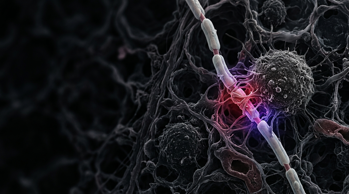
In mouse models of melanoma and breast cancer, researchers found metastatic cells next to vagal sensory nerve fibers in the lung. Both cell surfaces carried NINJ1, a protein involved in nerve injury and repair. NINJ1 on the tumor cell bound to NINJ1 on the neuron.
Contact Stabilized Beta-Catenin
The contact trapped components of the tumor cell’s beta-catenin destruction complex, including AXIN1 and DVL1. Beta-catenin remained active and increased the expression of IGF1R and IGF2R, two receptors that respond to insulin-like growth factors.
The Tumor Cells Then Used Local Growth Signals
Macrophages and fibroblasts in the lung supplied IGF-1 and IGF-2. The NINJ1 contact prepared metastatic cells to respond to those local signals, connecting a physical encounter with the nerve to a growth program inside the tumor cell.
Blocking NINJ1 Reduced Lung Metastasis
Surgical, genetic, and pharmacological disruption of vagal sensory signaling reduced tumor burden in the reported mouse experiments. Removing NINJ1 from the cancer cells or blocking it with antibodies also reduced metastatic growth.
The Pathway Is an Early Therapeutic Target
The work is a preprint based on animal models. It identifies a specific neuro-metastatic pathway and a protein that can be blocked experimentally. Human studies will determine whether the same contact helps metastatic cells colonize the lung in patients.