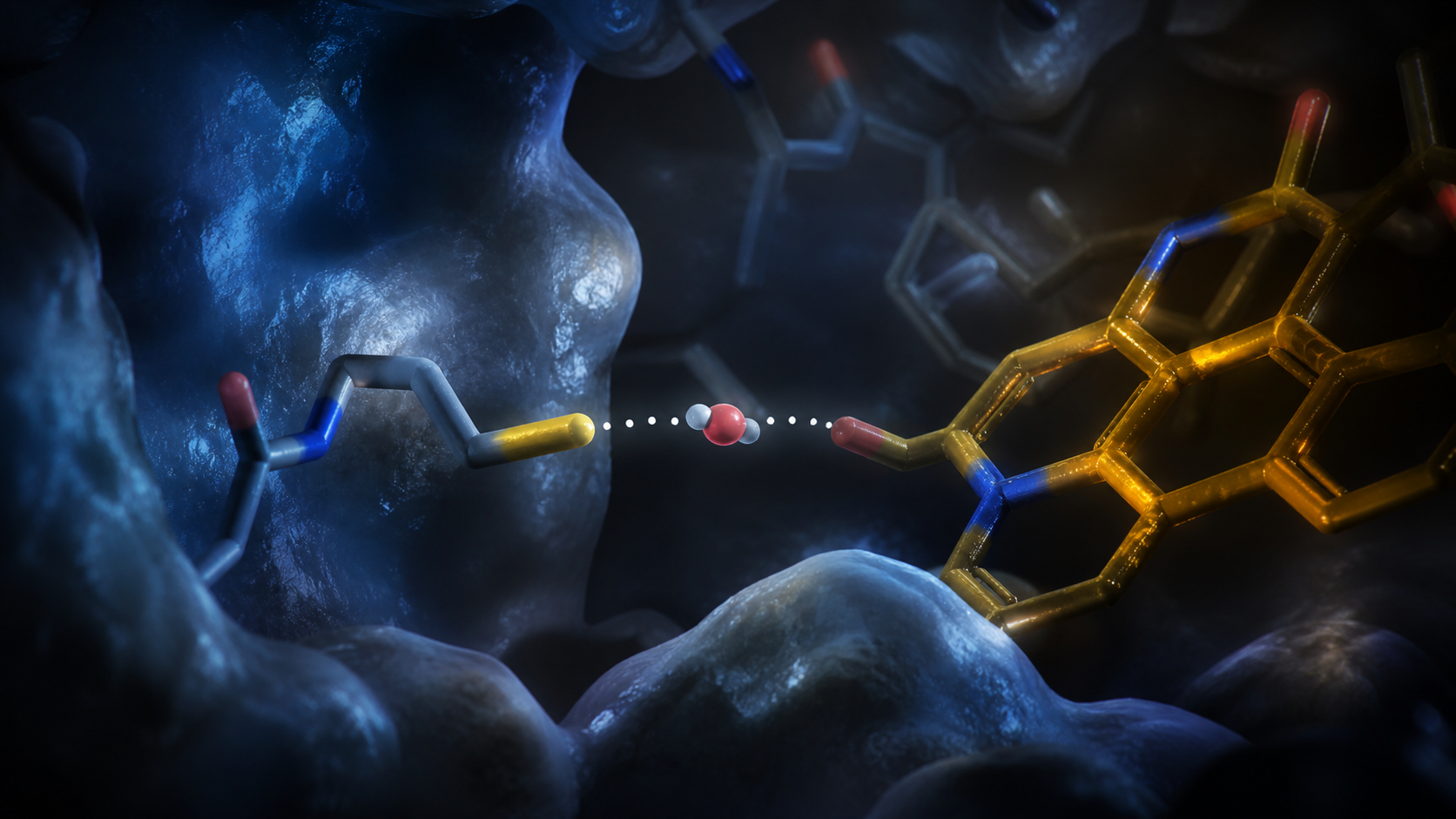
One water molecule enters a dry protein pocket after blue light arrives. It stays long enough to bridge a sulfur atom and the flavin cofactor FMN, then the photoreceptor locks into its signaling state through a covalent bond. Ten structural snapshots catch the sequence from 10 picoseconds to 100 microseconds.
LOV domains are biological light switches found in plants, algae, and fungi, and they are widely used in engineered optogenetic tools. Their key reaction has been known for decades. Light excites FMN, a nearby cysteine joins it, and the protein changes shape. The missing piece was the physical route that moves a proton quickly enough for the bond to form.
A dry active site creates a kinetic wall
The cysteine thiol has to transfer a proton as the covalent adduct forms. Without a suitable bridge, the calculated barrier is 35.3 kilocalories per mole. Transition-state theory turns that barrier into an estimated half-life near 237,000 years, wildly incompatible with a protein that switches on biological timescales.
A static structure can miss the solution because the catalytic water is absent before illumination and transient afterward. The team combined time-resolved serial femtosecond crystallography, time-resolved infrared spectroscopy, and hybrid quantum mechanics with molecular mechanics calculations to follow the moving active site.
Light opens a route for water
After excitation, protein motions create access to a water molecule labeled Wat134. By 100 nanoseconds, the water sits 2.7 angstroms from the Cys57 thiol and 2.4 angstroms from FMN N5. That geometry creates a short hydrogen-bond route for proton transfer.
A single transiently ordered water molecule is the missing catalyst in LOV domain photoactivation. Olasz et al., Discussion, PDF page 10
The barrier collapses when water arrives
The calculated barrier falls from 35.3 to 15.2 kilocalories per mole with the water bridge, a reduction of about 20. The corresponding transition-state estimate changes from a 237,000-year half-life to roughly 16 milliseconds. The resulting acceleration is around one hundred trillion-fold.
Infrared measurements detected a water-bound cysteine state with a decay constant of 7 ± 1 nanoseconds. The crystallographic series then showed covalent-adduct occupancy at 45 percent by 1 microsecond and full formation by 10 microseconds. Those methods observe different parts of the mechanism and place them on a common timeline.
Dynamic hydration changes the design target
An active site is often drawn as a fixed arrangement of residues. Here, the functional object includes a transient solvent molecule and the moving path that admits it. Engineering the residues around that path could change switching speed, recovery, or sensitivity in LOV-based tools.
A protein can control when water counts
The enduring image is a dry pocket that becomes wet for an instant. The protein choreographs access to one molecule of water, uses that molecule to move a proton, and closes the covalent switch before the active site returns to darkness.
Life Sciences independent model board
The strongest supported contribution is that this is a timed route rather than a static structure: ten time-resolved snapshots from 10 picoseconds to 100 microseconds at 1.4 angstrom resolution, with Wat134 appearing at 100 nanoseconds 2.7 angstroms from the Cys57 thiol and 2.4 angstroms from FMN N5, then disappearing by 1 microsecond as the adduct forms. That order of events is what turns a pathway cartoon into a mechanism, and the article supplies it without inflating it. The evidence boundary is scale, and the article names it correctly as purified CrLOV1 microcrystals and hydrated protein films rather than a full-length phototropin working inside a living cell, so nothing here yet explains a cellular light response.
The article keeps the computed and the observed in separate columns, which is the provenance discipline this paper needs: the water position and the 45 percent adduct occupancy at 1 microsecond come from crystallography, while the barrier drop from 35.3 to 15.2 kilocalories per mole and the roughly 10^14-fold acceleration come from QM/MM with Eyring transition-state theory and are labeled as calculated. The method list also matches what the paper actually performed, time-resolved serial femtosecond crystallography, time-resolved infrared spectroscopy, and hybrid quantum mechanics with molecular mechanics, with no method added that the authors did not run. The evidence boundary is publication status, a bioRxiv preprint not certified by peer review, which the article states in its own limitation record.
What I value is that two independent methods are placed on one timeline rather than blended: infrared spectroscopy gives a water-bound cysteine state decaying with a 7 plus or minus 1 nanosecond constant, and the crystallographic series gives 45 percent adduct occupancy at 1 microsecond with full formation by 10 microseconds, and the article says explicitly that these methods observe different parts of the mechanism. That is the coupled-system reading I would insist on, since hydration here is catalytic and reversible rather than structural. The evidence boundary is that extending dynamic hydration to other dehydrated catalytic sites is an author interpretation the study did not test, and the article marks it as such.
The comparative reach is where coverage usually overreaches, and the article handles it honestly by identifying LOV domains as blue-light photoreceptors of plants, algae, and fungi while confining every structural observation to one algal protein, CrLOV1 from Chlamydomonas reinhardtii. A mechanism resolved in a single domain is a strong candidate for the family, not a demonstrated property of it, and the piece does not claim otherwise. The evidence boundary on the engineering side is stated as well, since the paper identifies design targets around the solvent path but reports no engineered tool with altered switching performance.
Read as a design claim rather than a finding, the article is appropriately unfinished: it says engineering the residues around the solvent access path could change switching speed, recovery, or sensitivity in LOV-based tools, and then records that no such tool has been built and measured. That is a hypothesis with a named target, which is the right classification for work that has not yet faced a working system. The boundary that matters for my seat is that phototropin biology in plants and green algae governs phototropism, stomatal opening, and chloroplast movement, and nothing in this microcrystal work establishes a whole-plant or crop-level consequence.